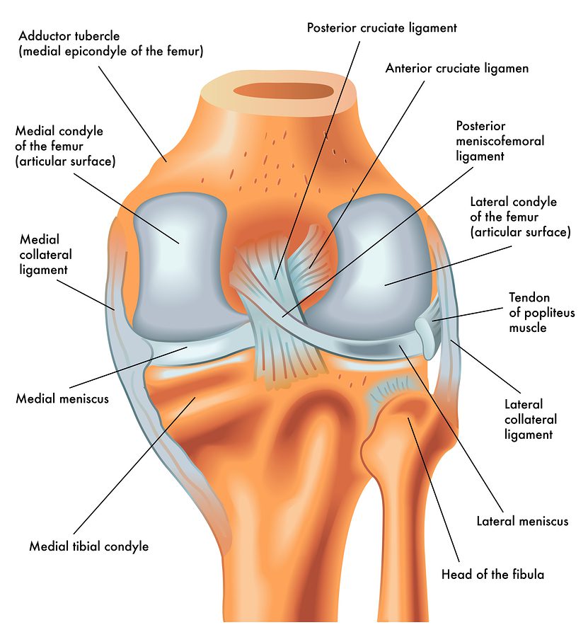
Ligaments are tough bands of tissue that connect the bones in the body. There are four ligaments in the knee that are prone to injury namely Anterior Cruciate Ligament (ACL), Posterior Cruciate Ligament (PCL), Medial Collateral Ligament (MCL), and Lateral Collateral Ligament (LCL).
Anterior Cruciate Ligament (ACL) is the most commonly injured knee ligament. It connects the thigh bone to the shin bone. An anterior cruciate ligament (ACL) tear is an injury to the knee commonly affecting soccer players, basketball players, skiers, gymnasts, and other athletes. About 70% of ACL tears are the result of non-contact injuries; 30% are the result of direct contact (player-to-player, player-to-object). Women are 4 times more likely than men to experience an ACL tear. It can tear due to twisting the knee while keeping the foot planted on the ground, sudden stop while running, sudden shift of weight from one leg to the other, jumping and landing on an extended (straightened) knee, stretching the knee farther than one should and experiencing a direct hit to the knee.
Posterior Cruciate Ligament (PCL) also links the thigh bone to the shin bone in the knee. The PCL is one of the four ligaments in the knee and is the ligament that prevents the tibia (shin bone) from sliding too far backward. Along with the ACL which keeps the tibia from sliding too far forward, the PCL helps to maintain the tibia in position below the femur (thigh bone). PCL injuries account for about 20% of knee ligament injuries. The most common PCL injury is often referred to as a “dashboard injury.” This injury is often seen in car accidents when the shin forcefully strikes the dashboard. The knee is in the bent position, and an impact forcefully strikes the shin backward. This injury can also occur when an athlete falls on the front of their knee which hyperflexes the knee (bends all the way back), with the foot held pointing downwards. Both scenarios stress the PCL and if the force is high enough, a PCL tear will result. Symptoms of a PCL tear are knee pain, swelling, and decreased motion. Some may say that their knee “popped” or gave out.
Medial Collateral Ligament (MCL) links the thigh bone to the shin bone on the inside of the knee. The MCL is another one of the four ligaments in the knee. The MCL is on the inside of the knee joint and spans the distance from the end of the femur (thigh bone) to the top of the tibia (shin bone). The MCL resists widening of the inside of the joint, or prevents “opening-up” of the knee. The MCL is usually injured when the outside of the knee joint is struck, causing the outside of the knee to buckle and the inside to widen. When the MCL is stretched too far, it may tear. An MCL injury may be an isolated injury or it may be part of a complex injury to the knee. Other ligaments, most commonly the ACL or the meniscus may be torn along with an MCL injury. MCL injury pain is usually focused directly over the ligament. Bruising and generalised joint swelling are common a few days after the injury. In some cases, some may feel as though their knee may ‘give out’ or buckle. Symptoms of an MCL injury tend to correlate with the extent of the injury.
Lateral Collateral Ligament (LCL) connects the thigh bone to the fibula, the smaller bone of the lower leg on the outer side of the knee. The LCL is on the outside of the knee, and connects the end of the thigh bone (the femur) to the top of the smaller shin bone (fibula). The LCL helps to prevent excessive side-to-side movement of the knee joint. When the LCL is torn, the knee joint may move too far side-to-side when stressed. The LCL is most commonly torn during sports activities or traumatic injuries (falls).
Physical therapy is vital in assisting in the care of ligament injuries. Non-surgically managed ligament injuries are often treated with relative rest, physical therapy, bracing, and a progressive exercise program. Physical therapy will be helpful in dealing with early pain and inflammation by using manual therapy and modalities. Light intensity exercises can be started almost immediately and progressed as tolerance allows. Sport and activity-specific work will begin as soon as appropriate.
For surgical cases, physical therapy plays an even more important role. Most surgeries to repair ligaments in the knee require extensive rehabilitation before and after the surgery, which is coordinated by a physical therapist. Managing swelling and pain, and also assisting with a gradual progressive exercise program in accordance with the surgeon’s post-operative protocol is the main job of the physical therapist early on. Increased functional and sport-specific training is started as soon as the patient’s progress allows. If in doubt, seek professional advice.
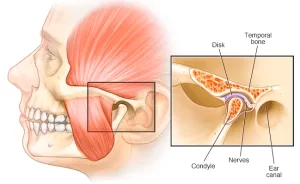
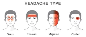
Check out our popular articles: Diastasis Recti, Tight Back Muscles, Irritable Bowel Syndrome (IBS), Temporomandibular Joint (TMJ) Dysfunction, Tennis Elbow, Wrist Tendon Injury, Sciatica, Whiplash, Hernia, Herniated Disc (Slipped Disc).